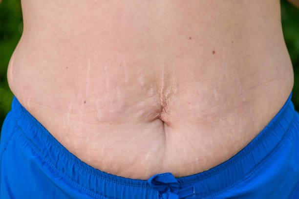
A CASE OF HUGE ANTERIOR ABDOMINAL WALL SWELLING MIMICING A TUMOUR – A DIAGNOSTIC DILEMMA – A RARE INTERESTING CASE

CLINICAL HISTORY
Here we report a case of 43 year old man presenting with huge swelling over left side
anterior abdominal wall in subcutaneous region following RTA for the past 1 year
which was clinically diagnosed as ? Haematoma /? Desmoid tumour. Cyst was
removed intoto and sent for HPE.
HISTOPATHOLOGICAL EVALUATION
GROSS DETAIL (HP 1006/24) – A huge cystic mass measuring 16.0 x 14.0 x 8.0 cm.
External surface appears grey brown attached with fat. Cut surface around 150 ml
clear brownish uid left out and shows multiloculated cysts. Cyst wall markedly
thickened. Inner surface irregular. No solid areas.
MICROSCOPY – Mul
tiple bits taken form cyst wall. Thickened foci of cyst wall show
parasites comprising of involuted scolex with hyaline degeneration and degenerated
larval form with duct like invagination lined by double layered eosinophilic membrane
surrounded by extensive foreign body giant cell granuomatous inammatory reaction.
Adjacent area shows evidence of resolving haematoma.
FINAL DIAGNOSIS: PARASITIC CYST – CYSTICEROSIS – ANTERIOR ABDOMINAL
WALL, SUBCUTANEOUS REGION.
DISCUSSION – Cysticerosis caused by infestation with larva of Taenia solium.
Transmission occurs by contaminated water or raw vegetables or pork infested with
larava.
Isolated cysticercosis of anterior abdominal wall without parasitosis of central
nervous system is very rare and may mimic a tumour leading to diagnostic and
therapeutic dilemma. Histopathological examination plays a important role in
making definite diagnosis. Laboratory tests usually show eosinophilia in the blood.
Serological tests have low sensitivity for solid cyst. FNAC and biopsy helpful for the
definitive diagnosis by demonstrating scolex.
Isolated soft tissue cysticerosis involvement has been used a marker of
neurocysticercosis. Therefore evaluation of cerebral and ocular involvement is
recommended. In our case we excluded the involvement of CNS and ocular region.
Surgical excision of the Isolated soft tissue Cysticercosis is sufficient if coexisting
CNS or ocular involvement has been ruled out. Medical treatment is by taking
Albendazole. If diagnosed correctly, it can be treated medically without the need for
surgery.
CONSULSION: Clinical diagnosis often difficult due to nonspecific manifestations of
cysticerosis of abdominal wall. However clinical diagnosis should be considered in
differential diagnosis especially in endemic regions and areas consuming pork.


